Complete Dental Services in Ardmore, OK
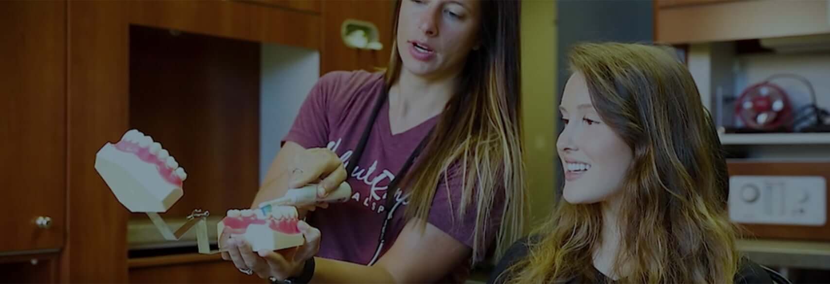
At Walnut Ranch Dental Spa, we never create cookie-cutter treatment plans—your comprehensive plan is made for you and only you. Our team is here to help you obtain a smile you can feel confident in, and we believe there’s something for everyone at our office! When you trust us with your oral health, you’ll be getting personalized care from a team with your interests at heart. Whether you’re coming to us for preventive, restorative, or cosmetic dental work, our team will be happy to work with you until we help you achieve your unique smile goals. Our Ardmore dental office never puts in the bare minimum and calls it a day; we are truly committed to providing high-quality care for all of our valued patients. To learn more about our services or schedule your next visit, please contact us!
Contact Us
Preventive Dental Services
Preventing dental issues before they develop is always preferable. Our preventive treatments are designed to help keep tooth decay, gum disease, and dental trauma at bay. We always recommend scheduling routine dental visits with our team at least twice per year to receive a thorough dental exam and professional teeth cleaning to keep your teeth and gums in tip-top shape. During these visits, we can also recommend additional preventive measures that we believe will help your smile. Our preventive dental services include:
- Routine Cleanings & Exams
- Dental Sealants
- Fluoride Treatments
- Oral Cancer Screenings
- Athletic Mouthguards
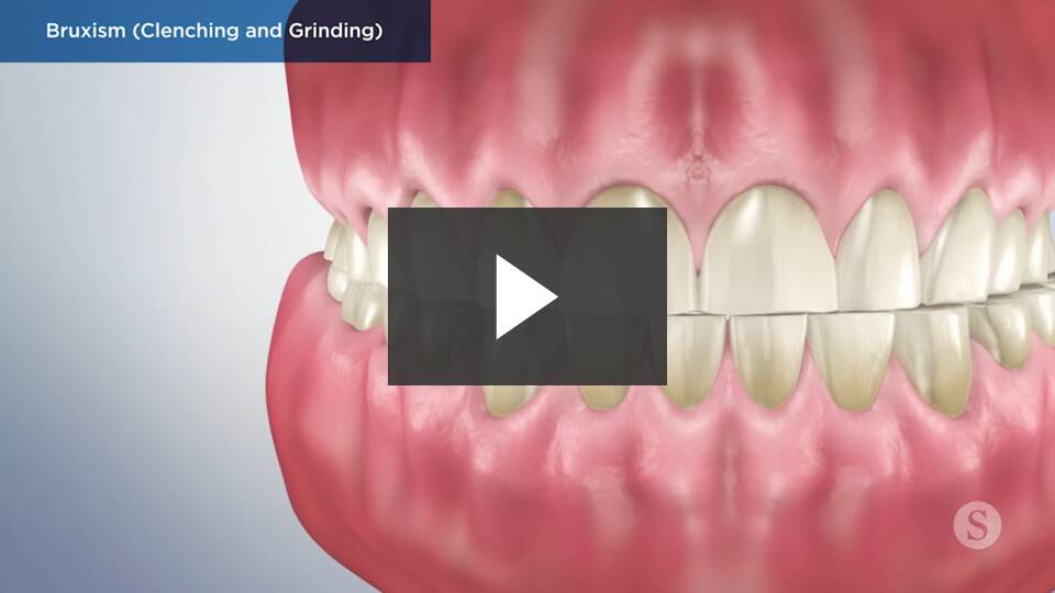
- Nightguards (For bruxism)
- TMJ/TMD Therapy

Restorative Dental Care
Restorative dental work can sometimes become necessary to repair your teeth and gums and improve your oral health. Walnut Ranch Dental Spa is committed to practicing conservative dentistry, which means we never recommend treatments that we don’t believe will ultimately benefit the patient. Our team wants to utilize the least invasive treatment methods possible to provide our patients with exceptional results. Our restorative services include:
- Tooth-Colored Dental Fillings
- Dental Crowns & Bridges
- Root Canal Therapy
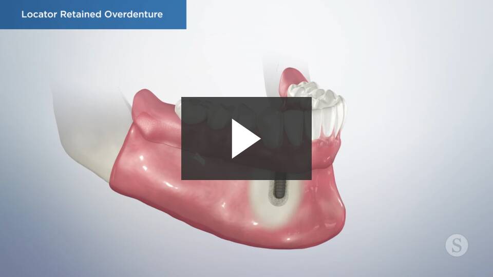
- Full & Partial Dentures
- Implant-Supported Dentures
- Dental Implants
- Dental Extractions
- Wisdom Teeth Removal
- Bone Graft Procedures

Cosmetic Dental Care
Cosmetic dental treatments are designed to enhance the natural beauty of teeth to help patients attain their dream smiles. Whether you’re looking to boost the brightness of your teeth or you’re interested in a complete smile makeover, our team can help. Dentistry is not just about improving your oral health; we want you to feel very confident about the appearance of your smile as well! Not sure which treatment option is right for you? Feel free to schedule a cosmetic consultation with our Ardmore dental team, and we’ll be happy to assist you. Our cosmetic services include:
- Professional Teeth Whitening
- Traditional Veneers
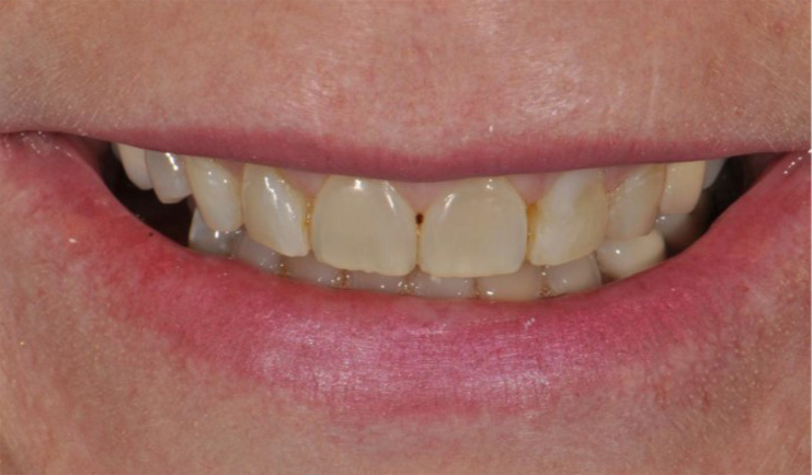
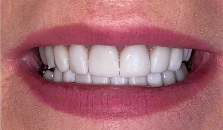
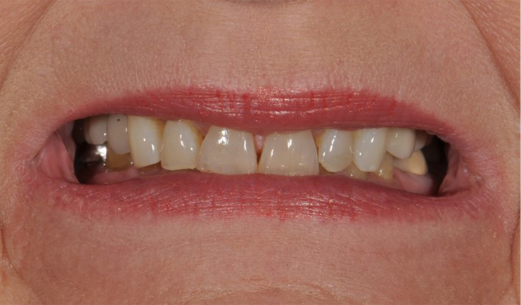
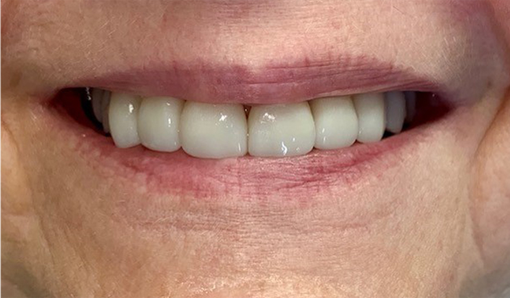
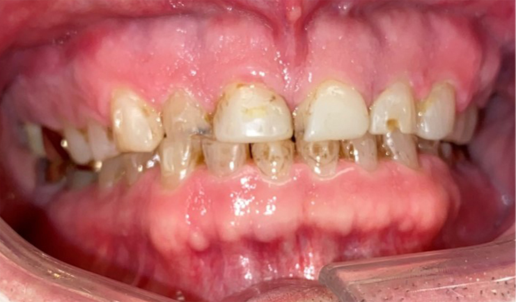
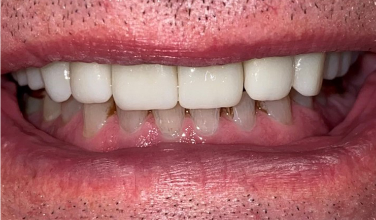
Before & After Cosmetic Treatment
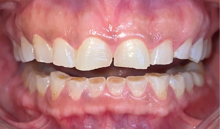
Before
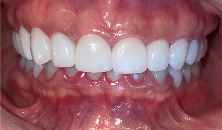
After
Before
After
Before
After
Before
After
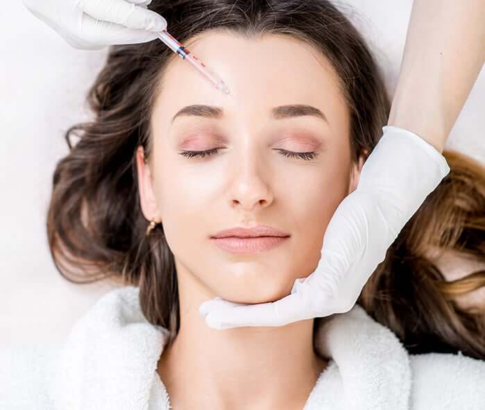
Dermal Filler
Dental spas are the perfect choice for patients who want to receive additional cosmetic treatments, like Botox and dermal filler, in addition to their routine dental care. Our filler services help patients achieve firmer, more youthful-looking skin, and they are often referred to as “lunchtime treatments” due to how quickly they can be completed. Dermal filler can be used to replace lost facial volume, plump thin lips, and even reduce the appearance of scars. Our team would be happy to go over our dermal filler options with you to help you complete your cosmetic goals!

Dental Sedation
Dental sedation can be helpful for patients who experience dental anxiety and patients who are undergoing lengthy restorative procedures. Our practice offers a few different choices for dental sedation, and we would be happy to review your current health status, medical history, treatment plan, and preferences to determine the ideal sedation option for you. Our safe and effective dental sedation options include:
- Nitrous Oxide
- Oral Conscious Sedation
- IV Sedation
- Oraverse